Fascination Medical Imaging: We make the invisible visible
Klaas Prüssmann is professor of Bioimaging and the head of the Institute for Biomedical Engineering (IBT). In this interview, he speaks about his mission to generate images from inside the human body and how he came to ETH Zurich rather accidentally.
Professor Prüssmann, can you explain your research focus?
We create images for medicine, particularly from inside the human body. These images are not only crucial for patient examinations but also for fundamental research. Our goal is to extract as much information as possible from within the body and represent it visually. To achieve this, we develop non-invasive techniques that do not harm the human body.
Why are images so important in your research?
Images have been an essential part of human perception for millions of years. The saying "a picture is worth a thousand words" is particularly true, as a signifcant portion of our brain is specialised in processing visual stimuli. Therefore, we use imaging techniques to visualise complex information. Images provide a spatially resolved representation of the world that is intuitively understandable. This is especially crucial in medicine, where precise and comprehensible depictions of the body’s interior are essential.
What physical principles do you use in your research?
Our work primarily relies on magnetic interactions, specifically nuclear magnetic resonance. Magnetism is advantageous because the human body is only slightly magnetic. For magnetic fields, the body is quite transparent – a key requirement for looking inside. If the body were strongly magnetic, too many interactions would overlap, making signals difficult to interpret. This is also why imaging through electric fields is less practical. The body itself is quite electric and electrically active, which would lead to confusion and potential harm when using electric fields.
How did you get into magnetic resonance research?
It happened rather by chance. In 1994, I was deciding on a topic for my diploma thesis. In Bonn, where I was studying physics and medicine, the thesis research would have taken a year. At a party, a friend told me about ETH Zurich, where supposedly it could be done in half a year. That sounded tempting, so the next morning I travelled to Zurich. I wandered around the Hönggerberg campus for a day, to no avail. Back down in the valley, I remembered a Zurich textbook on magnetic resonance and spontaneously decided to visited the author, Peter Bösiger, the head of the Institute for Biomedical Engineering, in his office. He was there and had a long list of fascinating topics. That’s how I ended up in Zurich. In the end, my thesis still took a year – it was still subject to the Bonn curriculum.
“How did I get into magnetic resonance research? My diploma thesis was on magnetic resonance. Since then, this field has always proved to be exciting and full of opportunities for me.”Prof. Klaas P. Prüssmann
What impact does your research have on society?
Our research has a direct impact on medicine, especially in diagnostics, which grows more sensitive and differential with more image information. We also strive to reduce cost, simplify examinations, and improve device efficiency. Moreover, we train experts who disseminate their knowledge into society. And our work also influences the economy. A notable example is our spin-off company, Skope, which develops measurement technology and manufactures key components of MR devices. Today, it belongs to a large global company. These advancements contribute to improving medical care and making it more accessible.
What are the biggest challenges in your field of research?
One major challenge is to make previously invisible constituents of the body visible. Traditional MRI mainly visualises fluids like water, but we aim to also image solid structures and macromolecules such as myelin and collagen. Myelin is the insulating layer around nerve fibres and plays a crucial role in the brain. Its degradation is associated with severe diseases. Collagen is a protein found in many tissues and is essential for their mechanical stability. Visualising these structures could lead to significant diagnostic advances.
Can you elaborate on the technical challenges and solutions in your research?
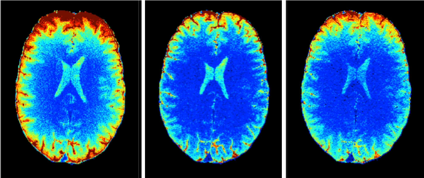
One persistent challenge is the unavoidable movement of patients during imaging. The higher the resolution, the more sensitive the image quality is to even the smallest positional changes. We are working on detecting and immediately correcting for movement during examinations by co-rotating the critical magnetic fields. This requires advanced sensor technology and control engineering. These two areas of electrical engineering also help us overcome the limited fidelity of MRI device components. Through sensor technology, modelling, and rapid computation, we are expanding the capabilities of MRI.
What role does artificial intelligence (AI) play in your research?
AI is an important tool, particularly for pattern recognition in our images. It can help detect diseases at an early stage and improve data analysis. However, everything starts with physical interaction and data acquisition before AI can be useful. AI can process vast amounts of data in little time and extract valuable information. This has the potential to revolutionize diagnostics by assisting radiologists and increasing efficiency. However, there are challenges, such as integrating new image contrasts and adapting medical practice to these emerging technologies.
You teach at both ETH and the University of Zurich – how do you experience the differences between the two institutions?
The collaboration, in our case between D-ITET and the Faculty of Medicine, is close and productive. The two sides complement each other perfectly, and there is much to be done together. Each professorship at IBT has a "leading house" (either ETH Zurich or the University of Zurich), but cooperation within the institute is seamless. There are some procedural differences between the universities, but these are manageable, and the benefits of collaboration far outweigh the challenges.
What are your visions for the future of your research?
Physics still offers many opportunities to enhance the information content of MRI and other magnetically detected data, such as magnetoencephalography (MEG). The sheer volume of data is already pushing human observers to their limits. However, a new, wide playing field emerges when richer raw data is converted into added medical value with the help of machine analysis. I expect that the interplay with machine learning will give another boost to the utility of our work. The new GLC building, where MRI and MEG systems are housed side by side, provides us with ideal conditions for this research. After years of planning and construction, the completion of the GLC has propelled us forward significantly.
Are you looking for new scientific staff?
With our move into the GLC, we have the capacity to grow. Our research group is highly international and mainly consists of graduates in electrical engineering, physics, and biomedical engineering. Students from related fields such as computer science and mechanical engineering also join us occasionally. We are definitely looking to expand this year and welcome enquiries from interested candidates.